Participant choice
This examine was carried out in accordance with the tenets of the Declaration of Helsinki. This examine was authorised by the Institutional Assessment Board of the Fukui College Hospital, Fukui, Japan. This examine was registered with the College Hospital Medical Data Community Scientific Trials Registry of Japan (identifier: College Hospital Medical Data Community 000046665, date of entry and registration: January 18, 2022). Individuals had been recruited between January 18, 2022, and March 14, 2022. Information had been collected from 16 eyes of 16 wholesome adults. Written consent was obtained from all contributors after an ample knowledgeable consent. Volunteers who had been handled with glaucoma medicines, with allergic conjunctivitis, or with different ocular illnesses had been excluded from the examine. Volunteers who had been pregnant or lactating or had the opportunity of being pregnant in the course of the examine interval had been additionally excluded. Contact lens customers saved their lenses off in the course of the examine.
Crossover drug administration
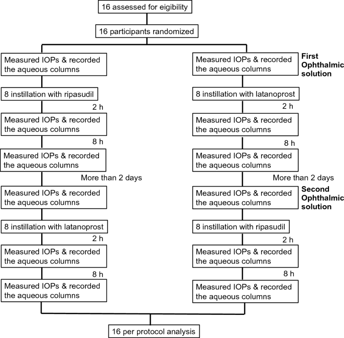
A 0.4% ripasudil hydrochloride hydrate ophthalmic resolution (GLANATEC®, Kowa, Nagoya, Japan) and a 0.005% latanoprost ophthalmic resolution (Xalatan®, Viatris, Tokyo, Japan) had been used on this examine. Wholesome volunteers had been randomized at a 1:1 ratio to one of many two crossover sequences to the instillation with ripasudil or latanoprost ophthalmic resolution. After a washout interval of greater than 2 days, they had been crossed over to the choice instillation (Fig. 1).
This examine was carried out in a double-blind method. A random quantity desk was used to find out the ophthalmic resolution that was first instilled to every volunteer. The investigator, who randomly assigned volunteers, instilled ophthalmic options with out letting them know which medication had been instilled. One other investigator measured IOPs with applanation tonometry and recorded the aqueous columns within the episcleral veins with a video seize system related to the slit-light microscope (hemoglobin video imaging) earlier than and a couple of and eight h after the instillation.
Measurement of aqueous column
The width of the aqueous column was decided in response to a earlier examine8. Briefly, we set a forty five× magnification of the slit-lamp microscope. A inexperienced filter (between 505 and 575 nm wavelengths) was used to visualise episcleral veins and aqueous columns. We used an iPhone 11 (Apple, Cupertino, CA) to report the video motion pictures. The iPhone 11 was related to the eyepiece a part of the slit lamp and set to 1.5× magnification. The pictures had been captured in 4K at 60 frames per second. Once we analyzed the captured photographs, we alternately skipped one body out for the 60 frames after which analyzed 30 frames per second. Photographs had been recorded 4 and 9 occasions to report steady aqueous move within the episcleral vein. We chosen the pictures obtained instantly after blinking as a result of the aqueous move tended to decelerate with out blinking. The captured photographs had been transformed to the Audio Video Interleave format, and the information had been utilized to ImageJ (accessible at http://imagej.nih.gov/ij/). The width of the aqueous column was analyzed utilizing the ImageJ software program. We used the StackReg plugin to compensate for digital camera shake in video. The width of the aqueous column within the episcleral vein was outlined as the gap between the minimal depth (Supplemental Fig. 1) due to the cross-sectional view of the vein. The diameter was measured in 30-frame slices by two impartial observers (MS and YS), and the common worth was calculated.
The move charge was additionally calculated utilizing the next components described within the earlier report8:
$$Rij(n) = E[(pij (t){text{-}}{upmu })(Pij( t + n){text{-}}{upmu })]/ sigma ^{land}{2}$$
the place Rij(n) denotes the autocorrelation perform worth for a pixel at place (i, j) at a body delay worth t, µ is the imply pixel worth within the segmented picture, and σ is the usual deviation of the pixel values within the phase.
Information assortment
The best eye was chosen because the precedence within the current examine. Aqueous columns within the episcleral veins had been continuously noticed within the inferonasal quadrant10. If no aqueous columns had been noticed, photographs of the aqueous columns had been recorded within the inferotemporal quadrant. If no aqueous columns had been current within the inferior quadrant, the left eye was chosen. Individuals’ information included age, intercourse, laterality of the attention, IOP, and vein location.
Main end result measures
The first end result measures had been the comparisons between ripasudil and latanoprost for the modifications within the aqueous column width after the instillation.
Secondary end result measures
The secondary end result measures had been as follows; the affiliation between the width of the aqueous column and the speed of IOP discount from the baseline IOP; the modifications of the move charge after the drug administration; the incidence of the opposed occasions in the course of the examine.
Statistical analyses
Information are offered as imply ± customary deviation. A comparability of the change within the aqueous column width between the 2 forms of ophthalmic options at 2 and eight h after the instillation was carried out utilizing a linear blended mannequin. The sequences, ophthalmic options, and durations had been included within the mannequin as fastened results. The contributors inside a sequence had been included within the mannequin as random results. Comparability of the change between baseline and a couple of or 8 h after instillation was carried out utilizing the Wilcoxon signed-rank take a look at. Statistical significance was set at p < 0.05.


